pathogen id,
-
C. difficile Latex Agglutination Kit

Clostridium difficile has a well documented role in pseudomembraneous colitis and diarrhea associated with antibiotics. Evidence has also implicated C. difficile in colitis not induced by antibiotics, exacerbations of chronic inflammatory bowel disease and postoperative diarrhoea. Many laboratories rely on the detection of C. difficile cytotoxin to diagnose infection. However, investigations have shown that the detection of faecal cytotoxin is not always consistent with active infection. This, plus the fact that some laboratories do not have the specialised facilities that are required to detect faecal cytotoxin, has highlighted the need for a test that is easy to use and gives rapid results for the identification of C. difficile. Use of Microgen® C. difficile allows identification of C. difficile from selective agar, within two minutes.
Traditional methods for identification of C. difficile includes GLC and cytotoxin detection. However, GLC is expensive in terms of time, equipment and cost per test and the link between presence of cytotoxin and symptoms is inconsistent. The test latex particles in Microgen® C. difficile are coated with antibodies specific for C. difficile somatic antigens, resulting in a highly specific test with a high predictive negative value. The flexibility of the test means that it can be used to test isolates directly form selective agar plates, such as CCFA.
-
Campylobacter Latex Agglutination Kit

Microgen Campylobacter Latex
A latex agglutination test for the presence of Campylobacter sppon solid media or in enrichment broths. These assays are designed to provide simple, one step identification / confirmation of Camplylobacter. Immediate results detect presence of Camplyobacter, can be used in clinical, food, environmental and water testing laboratories.
Benefits
- Results in 2 minutes
- Cost effective
- High sensitivity
- High specificity
- Accurate & reliable results
- User friendly procedure

Included in the kit:
- 2.5mls Test latex
- 2.5mls Control latex
- 0.5ml Positive control
- 5mls Isotonic Saline (foods) or 50mls GBS (clinical)
- disposable reaction cards
- disposable mixing sticks
Detects the Campylobacter genus, including these common thermophilic species:
- C. jejuni
- C.coli
- C.jejuni subsp doylei
- C.upsaliensis
- C.laridis
- C.fetus
Recommended media
- Preston Broth - for enrichment from foods or feces
- Modified CCDA Preston - plates for isolation from foods or feces
Food Method from Plates
- Dispense 1 drop of saline on to 2 wells of the reaction card
- Using a loop, take colonies of typical morphology and make a heavy suspension in each drop of saline
- Add 1 drop of test latex to one well and 1 drop of control latex to the other, mix using a mixing stick
- Rock the card gently for up to 2 minutes and then read results
Clinical Method from plates
- Place 50µl of Sample Diluent on to 2 wells of the reaction card
- Remove a typical looking colony from the plate using a loop and emulsify in Sample Diluent
- Add 1 drop of test latex to 1 well and 1 drop of control latex to the other, mix using a mixing stick
- Rock the slide for up to 2 minutes and examine for agglutination
Clinical Method from broths
- Place 50µl of enrichment broth on to 2 wells of the reaction card
- Add 50µl test latex to one of the wells and 50µl control latex to the other, mix using a mixing stick
- Rock the slide for up to 2 minutes and examine for agglutination
Method Direct on Feces
- Add 0.1g or 0.1ml fresh feces to 1ml Sample Diluent in a clean tube.
- Shake briefly then allow coarse particles to settle
- dispense 50µl of the supernatant on to 2 wells of a reaction card
- Add 1 drop of test latex to one well and 1 drop of control latex to the other, mix using a mixing stick
- Rock the slide for up to 2 minutes and examine for agglutination
Campylobacter and food poisoning
Campylobacter enteritis is a very common cause of bacterial food poisoning in the US. Most cases are sporadic and associated with the consumption of contaminated poultry. Sporadic infection has also been associated with contamination of milk by birds pecking through the tops of milk bottles. Outbreaks are usually associated with consumption of non-pasteurized milk or untreated water. The peak incidences occur during the summer, infants and young adults affected most and is more frequent in rural areas.
Latex technology:
- Selected C.jejuniserotypes are grown up
- Cells harvested and purified
- Purified cell prep used to vaccinate rabbits
- Resulting IgG is absorbed against potential cross reactors eg Citrobacter
- Resulting antisera used to coat latex particles
Latex Agglutination Principle
- Polyvalent antisera raised against selected C. jejuniserotypes antigens is used to coat latex particles.
- When mixed with a suspension of Campylobacter containing these antigens, the latex particles rapidly agglutinate forming visible clumps.
Expected Results
- Direct test of feces - Microgen's Campylobacter latex will detect Campylobacter sppin ~70% of fecal samples from acute phase disease, within the first 2-3 days post onset of symptoms
- Testing Cultures - Microgen's Campylobacter latex will detect growth in >90% of campylobacter positive 24 hour cultures, providing the method is followed. The latex will typically detect 100% of campylobacter positive cultures sampled at 48 hours.
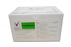
Campylobacter Latex Agglutination Kit
Storage: Store at 2-8°C Shelf Life: 1 year Detects the Campylobacter genus, including these common thermophilic species:
C. jejuni,
C.coli,
C.jejuni subsp doylei,
C.upsaliensis,
C.laridis,
C.fetus -
Gram Negative (GN) ID

GN-A kit

GN-B kit
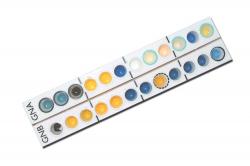
Identifies the complete range of Enterobacteriaceae and other non-fastidious, oxidase positive and negative, gram negative bacilli. The GN-A kit includes 60 microwell strips in foil pouches, frame for microwell strips, result forms, instructions for use and color chart for reading results. GN-A kit can be used separately from GN-B kit. GN-B kit includes 24 microwell strips in foil pouches and instructions for use. GN-B kit is designed to be used together with GN-A kit and not on its own. Identification software and reagents must be purchased separately.

Features list
- Simple to inoculate, handle and read
- Strip includes all tests in ISO and other standard methods
- No toxic reagents
- Results within 24 hours
- 100% sensitivity
- No MacFarland calibration
- Ability to isolate directly from chromogenic media
- No need for additional tests
- No assembly required
- No weak color changes
- Cost efficient
Easy to use:
- Select a single colony of the isolate to be identified
- Emulsify colony in 3ml saline (A) or 5ml saline (A+B
- Add 3-4 drops (100µL) of the suspension to each well of the strip(s)
- Overlay appropriate wells with mineral oil
- Incubate 18-24 hours at 35-37°C
- Next day add reagents
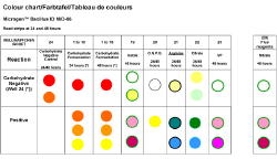
- Read and Record results using the color chart (provided) and Identification Software (purchased separately)
The GN-ID system comprises two separate, 12-substrate, microwell strips; the GN-A microwell strip and the GN-B microwell strip. The GN-A strip can be used separately and identifies the most commonly encountered, oxidase negative, non-fastidious, gram negative bacilli. GN-B is designed to be used in conjunction with GN-A; the combined strips identify the complete range of Enterobacteriaceae and other non-fastidious, oxidase positive and negative, gram negative bacilli. The wells in each microwell strip contain dehydrated biochemical substrates that are reconstituted with a saline suspension of the organism to be identified. If the substrate found in each well is metabolized by the organism, then a color change occurs during incubation or after the addition of specific reagents. Substrates are based on conventional biochemical methods used for identifying Enterobacteriaceae and other gram negative bacilli.
Procedure
A pure, 18-24 hour culture of the isolate to be identified must always be used. We suggest selecting a single colony from EZ-CHROM chromogenic media. An oxidase test must be performed on the isolate prior to strip inoculation. Oxidase positive organisms can only be identified when A and B strips are both inoculated. Emulsify a single colony from an 18-24 hour culture in sterile physiological saline (3 ml for strip A, 3-5 mls for A+B). Mix thoroughly. Add 3-4 drops (100µL) of the suspension to each well of the strip(s). After inoculation, overlay specified wells with 3-4 drops of mineral oil; these wells are highlighted with a black circle around the well to assist in adding oil to the correct wells. Seal the top of the microwell strip(s) with adhesive strip provided and incubate at 35-37oC for 18-24 hours. Remove adhesive strip and record all positive reaction with the aid of the color chart. Add Indole Kovacs, VP I & II, Nitrate A & B and TDA reagents to the appropriate wells as described in the Instructions for Use. Read and record results using the color chart and Identification Software (sold separately). For a complete explanation of procedure for use, please refer to the Instructions for Use, included with your kit.
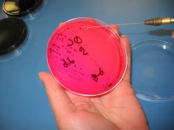
Step 1: Select a single colony
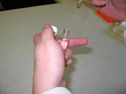
Step 2: Emulsify in saline solution
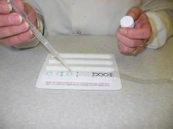
Step 3: Inoculate strip with solution
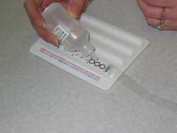
Step 4: Add mineral oil
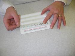
Step 5: Apply adhesive strip and incubate
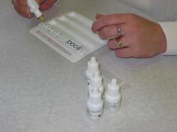
Step 6: Next day, add reagents
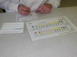
Step 7: Read and record results
The Microgen™ GN-A will identify the following:
- Acinetobacter spp.
- baumannii
- lwoffi
- A .haemolyticus
- Citrobacter spp.
- freundii
- diversus
- Enterobacter spp.
- aerogenes
- cloacae
- agglomerans
- E. gergoviae
- E. sakazakii
- Escherichia spp.
- E. coli
- E. coli-inactive
- E. vulneris
- Shigella spp.
- S. dysenteriae (Group A)
- S. flexneri (Group B)
- S. boydii (Group C)
- S. sonnei (Group D)
- Hafnia spp.
- alvei
- Klebsiella spp.
- K. pneumoniae
- K. oxytoca
- K. ozaenae
- K. rhinoscleromatis
- Morganella spp.
- M. morganii
- Proteus spp.
- P. mirabiliis
- P. vulneris
- Providencia spp.
- P. rettgeri
- P. stuartii
- P. alcalifaciens
- Salmonella spp.
- Salmonella Group I
- S. Typhi
- S. Cholerae-suis
- S. Paratyphi A
- Salmonella Group II
- Salmonella Group IIIa
- Salmonella Group IIIb
- Salmonella Group IV
- Salmonella Group V
- Salmonella Group VI
- Serratia spp.
- S. marcescens
- S. liquefaciens
- S. rubidaea
- Tatumella spp.
- T. ptyseos
- Yersinia spp.
- Y. enterocolitica
- Y. pseudotuberculosis
Species identified using the Microgen™ GN A + B Panel (OXIDASE NEGATIVE)
- Acinetobacter spp.
- baumannii
- lwoffi
- haemolyticus
- Budvicia spp.
- aquatica
- Buttiauxella spp.
- agrestis
- brennerae
- ferragutiae
- gaviniae
- B .izardi
- noackiae
- wamboldiae
- Cedecea spp.
- davisae
- lapagei
- neteri
- Cedecea sp 3
- Cedecea sp 5
- Citrobacter spp.
- freundii
- diversus
- C. amalonaticus
- C. farmeri
- C. youngae
- C. braakii
- C. werkmanii
- C. sedlakii
- C. rodentium
- Citrobacter sp 10
- Citrobacter sp11
- Edwardsiella spp.
- tarda
- tarda biogp1
- hoshinae
- ictaluri
- Enterobacter spp.
- aerogenes
- cloacae
- agglomerans
- gergoviae
- E. sakazakii
- E. taylorae (cancerogenus)
- E. amnigensis biogp1
- E. amnigensis biogp2
- E. asburiae
- E. hormaechei
- E. intermedium
- E. cancerogenus
- E. dissolvens
- E. nimipressuralis
- E. pyrinus
- Escherichia spp.
- E. coli
- E. coli-inactive (previously Alkalescens-Dispar Group)
- E. fergusonii
- E. hermannii
- E. vulneris
- E. blattae
- Shigella spp.
- S. dysenteriae (Group A)
- S. flexneri (Group B)
- S. boydii (Group C)
- S. sonnei (Group D)
- Ewingella spp.
- E. americana
- Hafnia spp.
- alvei
- alvei biogp1
- Klebsiella spp.
- K. pneumoniae
- K. oxytoca
- K. ornithinolytica
- K. planticola
- K. ozaenae
- K. rhinoscleromatis
- K. terrigena
- Kluyvera spp.
- K. ascorbata
- K. cryocrescens
- K. georgiana
- K. cochlea
- Leclercia spp.
- L. adecarboxylata
- Leminorella spp
- L.grimontii
- L.richardii
- Moellerella spp.
- M. wisconsensis
- Morganella spp.
- M. morganii
- M. morganii subsp morganii
- M. morganii biogp1
- M. morganii subsp siboni 1
- Obesumbacterium spp.
- O. proteus biogp2
- Pragia spp.
- P. fontium
- Pantoea spp.
- P. dispersa
- Photorhabdus spp.
- P. luminescens (25°C)
- Photorhabdus DNA Group 5
- Proteus spp.
- P. mirabilis
- P. vulgaris
- P. penneri
- P. myxofaciens
- Providencia spp.
- P. rettgeri
- P. stuartii
- P. alcalifaciens
- P. rustigianii
- P. heimbachae
- Rahnella spp.
- R. aquatilis
- Salmonella spp.
- Salmonella Group I
- S. Typhi
- S. Cholerae-suis
- S. Paratyphi A
- S. Gallinarum
- S. Pullorum
- Salmonella Group II
- Salmonella Group IIIa
- Salmonella Group IIIb
- Salmonella Group IV
- Salmonella Group V
- Salmonella Group VI
- Serratia spp.
- S. marcescens
- S. marcescens biogp1
- S. liquefaciens
- S. rubidaea
- S. odorifera biogp1
- S. odorifera biogp2
- S. plymuthica
- S. ficaria
- S. entomophila
- S. fonticola
- Tatumella spp.
- T. ptyeos
- Trabulsiella spp.
- T. guamensis
- Xenorhabdus spp.
- X. nematophilis (25°C)
- Xanthomonas spp.
- X.(S) maltophilia
- Yersinia spp.
- Y. enterocolitica
- Y. frederiksenii
- Y. intermedia
- Y. kristensenii
- Y. rohdei
- Y. aldovae
- Y. bercovieri
- Y. mollaretii
- Y. pestis
- Y. pseudotuberculosis
- Y. ruckeri
- Yokenella spp.
- Y. regensburgei
- Enteric gp58
- Enteric gp59
- Enteric gp60
- Enteric gp63
- Enteric gp64
- Enteric gp68
- Enteric gp69
Species identified using the Microgen™ GN A + B Panel (OXIDASE POSITIVE)
- Actinobacillus spp.
- Aeromonas spp.
- hydrophila
- veronii bio sobria
- veronii bio veronii
- caviae
- Alcaligenes spp.
- faecalis type 11
- faecalis
- xylosoxidans subsp xylos
- Burkholderia spp.
- cepacia
- pseudomallei
- Flavobacterium spp.
- F. meningosepticum
- F. odoratum
- F. breve
- F. indologenes
- Moraxella spp.
- Pasteurella spp.
- P. multocida
- P. haemolytica
- Plesiomonas spp.
- P. shigelloides
- Pseudomonas spp.
- Ps. aeruginosa
- Ps. fluorescens-25°C
- Ps. fluorescens-37°C
- Ps. putida
- Ps. stutzeri
- Ps. diminuta
- Shewanella spp.
- S. putrefaciens
- Vibrio spp.
- V. fluvialis
- V. furnissii
- V. mimicus
- V. vulnificus
- V. hollisae
- V. choleras
- V. parahaemolyticus
- V. alginolyticus
- V. cincinnatiensis
- V. damsela
- V. carchariae
- Weeksella spp.
- W. virosa
- W. zoohelcum
Gram Negative (GN) ID Storage The microwell strips are stable in the unopened foil pouches at 2-8oC until the expiry date stated. Once the foil pouch has been opened, unused microwell strips should be replaced into the pouch and the pouch resealed. Microwell strips in resealed pouches should be used within 14 days.
Shelf Life 1 year Well Substrates GN-A Strip Well 1- Lysine
Well 2- Ornithine
Well 3- H2S
Well 4- Gluclose
Well 5- Mannitol
Well 6- Xylose
Well 7- ONPG
Well 8- Indole
Well 9- Urease
Well 10- VP
Well 11- Citrate
Well 12- TDA
GN-B Strip Well 13- Gelatin
Well 14- Malonate
Well 15- Inositol
Well 16- Sorbitol
Well 17- Rhamnose
Well 18- Sucrose
Well 19- Lactose
Well 20- Arabinose
Well 21- Adonitol
Well 22- Raffinose
Well 23- Salicin
Well 24- Arginine
Reagents Indole Kovacs
TDA
Voges-Proskauer I
Voges-Proskauer II
Nitrate Reagent A
Nitrate Reagent B
Mineral Oil
-
Legionella Latex Agglutination Kit

L. pneumophila is the most common cause of Legionnaires Disease. At present 16 serogroups exist, of which L.. pneumophila serogroup 1 accounts for the majority of cases. In addition, a range of other species of Legionella have also been implicated as the causative agent of Legionnaires disease. Clinical and environmental awareness, and testing for Legionella, is rapidly increasing assisted by the introduction of several commercially available selective culture media.
However, none of the media are specific for L.. pneumophila serogroup 1 and not everything that grows on them is Legionella. Therefore a specific test is needed to quickly confirm the identification of isolates so that appropriate action can be taken.
Microgen® Legionella is an easy-to-use latex agglutination test for L. pneumophila serogroups 1 and 2-15 and now a range of non-L. pneumophila species which have been implicated as causes of diseases in humans. The kit provides specific results in under two minutes directly from a selective medium.
Conventional methods for identification of Legionella utilise specific antisera in standard agglutination or immunofluorescence tests. However, these methods lack sensitivity, are time consuming and can require specialised training and expensive equipment.
The test latexes used in Microgen® Legionella are coated with immunoglobulins which have been specially processed to minimise cross reactions with potentially cross reacting organisms, eliminating the need for a control latex, simplifying the procedure and increasing efficiency. Microgen® Legionella has recently been improved by the addition of a new latex reagent which identifies a range of non-L. pneumophila species of clinical significance with a high specificity and no cross-reactions with L. pneumophila.
Microgen® Legionella contains test latexes for both L. pneumophila serogroup 1 and 2-15, and now a latex reagent for the following additional species; L. micdadei, L. bozemanii 1 & II, L. dumoffii, L. longbeachae 1 & II, L. jordanis, L. gormanii, L. anisa and L. feelei in a convenient 50 test pack with a long shelf life to offer the most rapid, specific and sensitive test for Legionella currently available.
-
Listeria ID

Kit includes 20 microwell strips in individual foil pouches, 20 bottles of Listeria suspending medium, 1 bottle of red blood cells for haemolysin reaction, holding frame for test microwell strips, result forms and instructions for use. Identification software must be purchased separately.
Features:
- AOAC approved
- Inoculation is simple
- Identify from selective or non-selective media
- Final confirmation in 24 hours or less
- Unique haemolysis test included
- No additional testing needed
- No MacFarland standard
- No CAMP test
- No 24 hour blood plate
- No set-up
- No weak or confusing color changes
Easy to use:
- Select a single colony of the isolate to be identified
- Emulsify colony in the suspending medium provided
- Add 3- 4 drops (100µL) suspension to each well of the strip(s)
- Add 1 drop of haemolysin red blood cells to well 12 (shake vial)
- Incubate 18-24 hours at 35-37°C
- Read and Record results using software (purchased separately)
Species identified:
- L. monocytogenes
- L. innocua
- L. seeligeri
- L. ivanovii
- L. welshimeri
- L. grayi
AOAC-RI APPROVED!
Testing food and food ingredients for the presence of Listeria species, in particular L. monocytogenes, is one of the main pathogen tests performed in food testing microbiology laboratories. The Listeria ID strip enables laboratories to rapidly identify and confirm from a single colony isolated on selective or non-selective media. In 24 hours or less, Listeria ID will identify and confirm which of the 6 species of Listeria: L. monocytogenes, L. innocua, L. seeligeri, L. welshimeri, L. ivanoviiand L. grayi has been isolated.
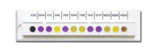
The Listeria ID system employs 12 standardized microwell biochemical substrates, including an in-well haemolysin test. The substrates are based on classical methods and include: Esculin, Mannitol, Xylose, Arabitol, Ribose, Rhamnose, Trehalose, Tagatose, Glu-1-Phos, Methyl-D-Glucoside, Methyl-D-Mannoside and Hemolysis. The inoculation and incubation of the substrate in each microwell causes a simple color change reaction, yellow is a positive reaction, purple is a negative reaction; there is no guess work involved.

Haemolysin Reaction
For convenience, an in-well haemolysin test is also included, eliminating the need for additional tests such as CAMP. As the detection of the haemolytic activity is fundamental to the identification of Listeria species (in particular L. monocytogenes and L. ivanovii) and the interpretation of this reaction is sometimes difficult, Microgen Bioproducts has developed a highly sensitive, easily interpreted and stable microwell haemolysin test. The haemolysin test is simply performed by adding 1 drop of the specially stabilized sheep red blood cells (included with kit) to the last well (well 12) of the Microgen Listeria ID test panel. If the organism being identified produces haemolysin, the red blood cells will rapidly be lysed and the cellular contents released into the suspending medium. The contents of the well will appear as a homogeneous red/ brown solution. Alternatively, if the organism being identified does not produce haemolysin, the stabilized red blood cells will remain intact. These cells will settle to the bottom of the microwell forming a distinct red layer with a clear supernatant.
Procedure
Using Listeria ID is simple and requires no set-up. Select a single colony of the isolate to be identified. The colony can be taken from selective or non-selective media, we suggest using BBL™ CHROMagar™ Listeria for presumptive identification. Emulsify colony in the suspending medium provided. Add 3- 4 drops (100µL) suspension to each well of the strip(s). Add 1 drop of red blood cells (provided) to the haemolysin test (well 12). Incubate for 18-24 hours at 35° – 37°C. Read and record results using the software program (sold separately). For a complete explanation of procedure for use, please refer to the Instructions for Use, included with your kit.

Emulsify single colony from selective or non-selective media in suspending solution

Inoculate strip with solution

Add red blood cells to hemolysin well and incubate
AOAC Approval: To establish the performance of the Listeria ID system, in an independent regulatory validation scheme, the product has undergone AOAC Research Institute (RI) Performance Method Tested (PMT) validations against the existing FDA/BAM method. The product was subjected to a range of validation studies including ruggedness, inclusively, exclusivity and performance in direct comparison to the FDA/BAM method. The performance of the product was also assessed using colonies taken from a range of selective, chromogenic and non-selective agars. The outcomes of all of these validation studies were reviewed by independent assessors and the product was granted Performance Method Status by the AOAC research Institute in September 2004. The results achieved in these rigorous validation studies should give confidence to routine food testing laboratories that this miniaturized biochemical identification system for Listeria speciation delivers a rapid method which performs to the highest standards in microbiological testing.
Listeria ID Technical Information Storage: The microwell strips are stable in the unopened foil pouches at 2-8°C until the expiry date stated. The Listeria suspending broth and haemolysin reagent should be stored at 2-8°C. The haemolysin reagent should be returned to 2-8°C immediately after use. Shelf Life: 1 year Species identified: - L. monocytogenes
- L. innocua
- L. seeligeri
- L. ivanovii
- L. welshimeri
- L. grayi
Well Substrates: - Well 1- Esculin
- Well 2- Mannitol
- Well 3- Xylose
- Well 4- Arabitol
- Well 5- Ribose
- Well 6- Rhamnose
- Well 7- Trehalose
- Well 8- Tagatose
- Well 9- Glu-1-Phos
- Well 10- Methyl-D-Glucoside
- Well 11- Methyl-D-Mannoside
- Well 12- Haemolysis
Reagents: None -
Listeria Latex Agglutination Kit

A latex agglutination presumptive test for the presence of Listeria spp to confirm colonies on agar plate media. Microgen Listeria latex may be used in conjunction with biochemical tests (Microgen Listeria-ID) for further species identification.
Benefits
- Results in 2 minutes
- Cost effective
- High sensitivity
- High specificity
- Accurate & reliable results
- User friendly procedure
Detects the Listeria genus, including these species:
- Listeria monocytogenes
- Listeria innocua
- Listeria welshimeri
- Listeria ivanovii
- Listeria seeligeri
- Listeria grayi
Included in the kit:
- 2.5mls Test latex
- 0.5ml Positive control
- 5mls Isotonic Saline
- disposable reaction cards
- disposable mixing sticks
Method from plates
- Place 1 drop of saline on to a well of the reaction card
- Remove a typical looking colony from the plate using a loop and emulsify in the drop of saline
- Rock the slide gently for 2mins to check for auto agglutination
- Add 1 drop of test latex to the suspension, mix using a mixing stick
- Rock the slide for up to 2 minutes and examine for agglutination
Interpretation
- Agglutination within 2 minutes is indicative of the presence of Listeria spp in the sample
- Absence of agglutination indicates that Listeria spp are not present in the sample
Latex Agglutination Principle
- Polyvalent antisera raised against the 4 Listeria flagella antigens is used to coat latex particles.
- When mixed with a suspension of Listeria containing these antigens, the latex particles rapidly agglutinate forming visible clumps
Latex technology:
- Producer strains carrying specific antigens are cultured at optimal temperature for flagella production
- isolates sub cultured on to Jameson plates to select most motile organisms
- strains cultivated in broth
- Supernatant discarded and cell deposit treated to harvest flagella
- flagella immunogen used to generate antisera
- antisera titres checked and blended
- blended antisera purified and then coated on latex particles
Listeria spp possess 4 major flagellar antigen types designated A, B, C and E. AllListeria spp possess one or more of A, B, or C butL. grayi only possess E. Antiserum used to coat the latex particles must contain all 4 flagella antigens in order to detect all 6 Listeria spp.
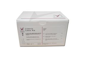
Technical Information for Listeria Latex Agglutination Kit Storage: Store at 2-8°C Shelf Life: 1 year Detects the Listeria genus,
including these species:- Listeria monocytogenes
- Listeria innocua
- Listeria welshimeri
- Listeria ivanovii
- Listeria seeligeri
- Listeria grayi
-
Microgen Bacillus ID

Microgen Bacillus ID is a miniaturized biochemical identification system designed to identify those Mesophilic Bacillus spp. and related genera associated with food and beverage spoilage and food poisoning. This identification system comprises 24 biochemical substrates SPECIFICALLY selected and formulated to provide accurate and efficient identification of Bacillus spp.
This product saves time and labor, while improving the accuracy and cost of identification of these organisms. The Microgen™ Bacillus ID employs the most up-to-date taxonomy. Each kit contains suspending medium and sufficient microwell strips for 20 identifications, holding tray, instructions, and reporting cards. Reagents and Identification Software are available separately.
Features:
- Small 24-well test system
- Simple, easy to use and read
- Final result in 48 hours
- Substrates specific for Bacillus
- Based on conventional methods
- Identifies all common species found in food and related samples
- Includes CARBOHYDRATE CONTROL well as reference for color interpretation
- Up-to-date taxonomy
Benefits:
- Confidence in results
- Quick to inoculate and simple to read
- Timely results
- Accuracy and security of reporting correct name
- Reduces errors
- Can cross reference with standard texts
- Cost effective
Easy to use and read:
- Select sufficient colonies from a purity plate
- Emulsify colonies in the suspending medium provided, equivalent to a MacFarland 2
- Inoculate each well of the strips
- Incubate 24 hours at 30°C
- Read and record carbohydrate, ONPG and citrate reactions
- Incubate further 24 hours at 30°C
- Add reagents then re-read strips and record results
- Analyze code with the Microgen Identification System Software
Bacillus Information:
- Clinical contaminationincludes operating rooms surgical dressings, and in pharmaceutical products.
- Food Spoilage:Bacillus spp. can be isolated from fruits, vegetable products, nuts, spices, cereals, milk and dairy products, meat and dried foods.
- Food Poisoning:Isolates of B.cereus, B.brevis, B.licheniformis, B.subtillis, and B.sphaericus have been found
- Milk and Milk Products:B.licheniformis is a common contaminant of dairy products. Paenibacillus spp., Brevibacillus spp. and Virgibacillus spp. are implicated in spoilage.
Species identified using the Microgen™ ID Bacillus
Bacillus species
- B. cereus group
- B. firmus
- B. badius
- B. laevolacticus
- B. coagulans
- B. lentus
- B. amyloliquefaciens
- B. subtilis
- B. circulans
- B. insolitus
- B. freudenreichii
- B. globisporus
- B. sphaericus
- B. thiaminolyticus
- B. pumilus
- B. licheniformis
- B. megaterium
Vergibacillus species
- V. pantothenticus
Paenibacillus species
- P. polymyxa
- P. macerans
- P. alvei
Brevibacillus species
- Br. brevis
- Br. laterosporus
Quick Guide to using the Microgen™ ID Bacillus ID Kit
- Step 1
Prior to the identification of any isolate it must be confirmed as belonging to the genus Bacillus.
- Gram positive bacillus
- Endospore forming
- Catalase positive
- Optimal growth temperature between 25 and 45°C i.e. Mesophilic. Isolates growing at < 25°C (Psychrophiles) or isolates growing at 45°C (Thermophiles) are not identified by this product.
- Step 2
-
Prepare a suspension equivalent to MacFarland No. 2 in the suspending medium provided. As some colonies may be very dry and difficult to emulsify to produce a homogeneous suspension it is recommended that a moist sterile swab be used to harvest colonies and re suspend. Additional mixing using a vortex mixer may be necessary. When using a swab to harvest growth, this should be done from a purity plate to ensure that mixed cultures are not created.
- Step 3
-
Peel back the adhesive sealing tape from each strip. Inoculate the microwells in both test strips with 3 drops (approx 100µl) of inoculum.
- Step 4
-
Overlay the Arginine well with Mineral oil.
- Step 5
-
Seal the strips. Incubate at 30ºC for 18 – 24 hours.
- Step 6
-
Read and record the results of all Sugar Fermentation tests PLUS the Citrate and Urease tests. Incubate at 30ºC for a further 18 – 24 hours.
- Step 7
-
Re read the results of all Sugar Fermentation tests. Add reagents (Indole, Nitrate, VP I & II).
NOTE: Occasionally some sugar fermentation tests may be positive after 18– 24 hours incubation but negative after 48 hours incubation. When the final Octal Code is calculated, all tests which showed a positive reaction after either 18 – 24 hours incubation or 48 hours incubation should be scored as POSITIVE. Only reactions negative at both readings should be scored as NEGATIVE.
- Step 8
-
Record all results on report form provided. Sum the accumulated positive test results from both the 24 and 48 hour readings.
- Step 9
-
Analyze the results using Microgen Identification System Software

This product saves time and labor, while improving the accuracy and cost of identification of these organisms. Each kit contains suspending medium and sufficient microwell strips for 20 identifications. Reagents and Identification Software are available separately. For further information, please contact Microbiology International at (800)396-4276.
For a complete explanation of procedure for use, please refer to the Instructions for Use, included with your kit.
Background Information
The genus Bacillus represents a large and diverse group of species including B. cereus which is well known as a causative agent of food poisoning. However, many other species of Bacillus and related genera have over the years also been implicated in outbreaks of food poisoning. Originally only 16 species of Bacillus were recognized. However, over the years additional species have been recognized and with the introduction of molecular methods, the genus Bacillus now includes 123 recognized species plus an additional 7 genera comprising closely related organisms are now recognized. Because these organisms are so widely distributed in the environment they are able to easily contaminate food ingredients and products. In addition, many of these organisms have been demonstrated to produce toxins capable of causing both diarrhea and vomiting. It is for this reason that the Microgen Bacillus ID includes not only some of the mesophilic Bacillus spp. but also some species belonging to Vergibacillus, Brevibacillus and Paenibacillus. All of the species included in this identification system have been isolated and/ or reported as causing food spoilage or food poisoning.
Bacillus species and food poisoning
Although B. cereus is the most common species of Bacillus associated with food poisoning, a range of other species have been implicated in foodborne illnesses. The two most commonly reported species are B. subtilis and B. licheniformis, these species being of concern to the food processing and canning industries as they are often implicated as causes of food spoilage producing potent proteolytic and saccarolytic enzymes. Some strains of B. licheniformis have been shown to produce toxins. In addition, a range of other species including B. pumilus, B. brevis and B. thuringiensis, have also been reported as being the sole species implicated in outbreaks of food poisoning. B. lentus, B. circulans, B. mycoides, B. polymyxa and B. laterosporus have also been demonstrated to produce toxins using cell cytotoxicity assays whilst some of these species have also been shown to produce positive RPLA and TECRA™ assays.
B. subtilis is the most frequently reported species associated with food poisoning other than B. cereus , being associated with outbreaks involving meat, pastry products, seafood and rice. Incubation periods varied from 1 – 4 hours, with recovery generally complete within 24 hours. Symptoms usually included nausea and vomiting, diarrhea was rarely reported. B. licheniformis associated food poisoning resembles that of C. perfringens, with an incubation period of 6 – 24 hours and symptoms including diarrhea and abdominal pain. In cases of food poisoning associated with B. licheniformis, high numbers of the bacteria 17 (106 cfu/gm) were found in the implicated food (often cooked meat and vegetable dishes) and in the feces of affected persons.
B. pumilus was implicated in 5 outbreaks of food poisoning between 1975 and 1986 in the UK , in each of these cases only a small number of individuals were involved, however in each case high numbers > 6 x 10⁶ organisms/ gm were isolated from the implicated foods in the absence of other recognized foodborne pathogens. Gilbert et. al. reported 4 instances of food poisoning in which B. brevis was the sole organism isolated in high numbers from foods implicated in food poisoning outbreaks. In any investigation of suspected food poisoning in which none of the normal primary pathogens such as Salmonella, Campylobacter, E. coli, C. perfringens and S. aureus are isolated Bacillus should always be considered as a potential cause. In such studies, particularly if the symptoms exhibited by patients include diarrhea, species other than B. cereus should always be considered.
Technical Information for Microgen™ Bacillus ID Storage: The microwell strips are stable in the unopened foil pouches at 2-8°C until the expiry date stated. The Bacillus suspending broth should be stored at 2-8°C. Shelf Life: 1 year Species Identified
Bacillus species
- B. cereus group
- B. firmus
- B. badius
- B. laevolacticus
- B. coagulans
- B. lentus
- B. amyloliquefaciens
- B. subtilis
- B. circulans
- B. insolitus
- B. freudenreichii
- B. globisporus
- B. sphaericus
- B. thiaminolyticus
- B. pumilus
- B. licheniformis
- B. megaterium
Vergibacillus species
- V. pantothenticus
Paenibacillus species
- P. polymyxa
- P. macerans
- P. alvei
Brevibacillus species
- Br. brevis
- Br. laterosporus
Well Substrates
- Well 1- Arabinose
- Well 2- Cellobiose
- Well 3- Inositol
- Well 4- Mannitol
- Well 5- Mannose
- Well 6- Raffinose
- Well 7- Rhamnose
- Well 8- Salicin
- Well 9- Sorbitol
- Well 10- Sucrose
- Well 11- Trehalose
- Well 12- Xylose
- Well 13- Adonitol
- Well 14- Galactose
- Well 15- Methyl-D-Mannoside
- Well 16- Methyl-D-Glucoside
- Well 17- Inulin
- Well 18- Melizitose
- Well 19- Indole
- Well 20- ONPG
- Well 21- Arginine
- Well 22- Citrate
- Well 23- VP
- Well 24- Nitrate
Reagents
- Indole Kovacs
- Voges-Proskauer I
- Voges-Proskauer II
- Nitrate Reagent A
- Nitrate Reagent B
- Mineral Oil
-
Salmonella Latex Agglutination Kit

Microgen Salmonella Latex
A latex agglutination confirmation test for the presence of Salmonella spp on solid media from clinical and food samples. Detects motile and non motile strains. Reduces testing delays associated with conventional methods. The Salmonella latex agglutination kit offers a rapid alternative to the traditional identification methods and detects a large range of Salmonella species including. S.typhimurium, S.enteritidis, S.virchow, S.newport, S.infantis, S.anatum, and various others belonging to serogroups B, C1, C2, C3, D1 and E1.
Benefits
- Results in 2 minutes
- Cost effective
- High sensitivity
- High specificity
- Accurate & reliable results
- User friendly procedure
Included in the kit:
- 2.5mls Test latex
- 0.5ml Positive control
- 5mls Isotonic Saline
- disposable reaction cards
- disposable mixing sticks
Method from plates
- place 1 drop of saline on to a well of the reaction card
- remove a typical looking colony from the plate using a loop and emulsify in the drop of saline
- rock the slide gently for 2mins to check for auto-agglutination
- add 1 drop of test latex to the suspension, mix using a mixing stick
- rock the slide for up to 2 minutes and examine for agglutination
Interpretation
- agglutination within 2 minutes is indicative of the presence of Salmonella spp in the sample
- absence of agglutination indicates that Salmonella spp are not present in the sample
Background
The genus Salmonella consists of >2000 serotypes. Can cause acute infectious enteritis frequently associated with food poisoning. Many Salmonella species infect domestic animals clinically or sub-clinically therefore many cases of Salmonella food poisoning originate from animal sources. In contrast S.typhiand S.paratyphionly infect humans and cause enteric fever. Salmonella are usually present in samples in low numbers therefore enrichment or selective media are used to promote their growth eg RV, SC, M- broth
Latex Agglutination Principle
- Polyvalent antisera raised against a variety of Salmonella flagella antigens is used to coat latex particles.
- When mixed with a suspension of Salmonella containing these antigens, the latex particles rapidly agglutinate forming visible clumps
- Microgen Salmonella Latex test detects the majority of common species including S. typhimuriumand S. enteritidis
- In addition, the non-motile S. gallinarumand S. pullorumare also detected as they retain the protein basal bodies which are the attachment points for flagella
Latex technology:
- Producer strains carrying specific antigens are cultured
- Jameson plates used to purify strain to single phase as strains can be present in 2 flagella phases
- strains cultivated to produce immunogen
- immunogen used to generate antisera
- antisera titres checked and blended
- blended antisera purified and then coated on latex particles
Technical Information for Salmonella Latex Agglutination Kit Storage: Store at 2-8°C Shelf Life: 1 year
-
Staph ID

Microgen® Staph-ID has been developed for the identification of commonly encountered Staphylococcus spp. isolated from clinical and veterinary samples. These bacteria, although commensal, are recognised as potential human pathogens e.g. S. aureus and S. epidermidis. Coagulase negative Staphylococcus spp. are common colonisers of prosthetic valves and in-dwelling catheters.
Staph ID Technical Information Storage: The microwell strips are stable in the unopened foil pouches at 2-8°C until the expiry date stated. The Staph suspending broth and haemolysin reagent should be stored at 2-8°C. The haemolysin reagent should be returned to 2-8°C immediately after use. Shelf Life: 1 year Species identified: - Staphylococcus spp.
- S. aureus subsp. aureus
- S. aureus subsp. anaerobius
- S. auricularis
- S. caprae
- S. capitis subsp. capitis
- S. capitis subsp. urealyticus
- S. carnosus
- S. chromogenes
- S. cohnii subsp. conhii
- S. cohnii subsp. urealyticum
- S. epidermidis
- S. haemolyticus
- S. hominis subsp. hominis
- S. hominis subsp. novobiosepticus
- S. hyicus
- S. intermedius
- S. lentus
- S. lugdenensis
- S. saccharolyticus
- S. saprophyticus
- S. schleiferi subsp. schleiferi
- S. schleiferi subsp. coagulans
- S. sciuri
- S. simulans
- S. warneri
- S. xylosus
- Kocuria spp.
- K. kristinae
- K. rosea
- K. carniphila
- Kytococcus spp.
- Ky. sedentarius
- Micrococcus spp.
- M. luteus
- M. lylae
-
Staph Latex Agglutination Kit

A rapid test for the identification and confirmation of Staphylococcus aureus colonies grown on selective solid media.
Two of the traditional diagnostic tests for S. aureus, the tube and slide coagulase tests, only detect the activity of a characteristic coagulase enzyme. However, these tests can be difficult to interpret and are liable to suffer from false positive results associated with non-specific factors frequently found in plasma. Non-specific coagulation due to other bacterial enzymes may also occur.
However, 99% of S. aureus isolates also produce detectable levels of protein A on their surface, a characteristic which can be used to differentiate S. aureus from other species of Staphylococci in which this protein is rare.
Microgen®Staph is a sensitive and specific latex agglutination for the identification of S. aureus which overcomes the problems associated with the traditional tests by detecting both coagulase and protein A, offering rapid and accurate identification of S. aureus in 2 minutes. Microgen®Staph contains all ancillary reagents needed to complete the test and is suitable for clinical, food and environmental laboratories.